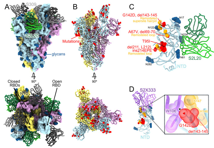
Fig. 1. CryoEM structure of the SARS-CoV-2 Omicron S trimer reveals a remodeling of the NTD antigenic supersite.
(A) Surface rendering in two orthogonal orientations of the Omicron S trimer with one open RBD bound to the S309 (grey) and S2L20 (green) Fabs shown as ribbons. The three S protomers are colored light blue, pink or gold. N-linked glycans are shown as dark blue surfaces. (B) Ribbon diagrams in two orthogonal orientations of the S trimer with one open RBD with Omicron residues mutated relative to Wuhan-Hu-1 shown as red spheres (except D614G which is not shown). (C) The S2L20-bound Omicron NTD with mutated, deleted, or inserted residues rendered or indicated as red spheres. Segments with notable structural changes are shown in orange and labeled. (D) Zoomed-in view of the Omicron NTD antigenic supersite overlaid with the S2X333 Fab (used here as an example of prototypical NTD neutralizing mAb (22)) highlighting the binding incompatibility; the modeled clash between S2X333 W106 and NTD G142D is indicated with an asterisk.

