Figure 4.
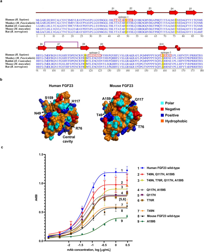
Conservation of antibody epitope in different species and reverse engineering of murine FGF23 to bind Burosumab. (a) The amino acid sequence alignment of FGF23 across binder (monkey and rabbit) and non-binder species (mouse and rat) is shown in a clustal omega format. The three distinct epitope regions (red box) were well conserved across, albeit with key species-specific differences. The residues that differentiate those binder species (monkey and rabbit) from non-binder species (mouse and rat) are shaded in yellow. The residues that make contact with Burosumab based on pose27 are colored in red. Secondary structures: β strand, α-helix, and a single disulfide formation between cysteine 93 and 113 are annotated as red arrow, red helix, and purple line, respectively. (b) The four putative cross-species determining residues: N49, R76, H117, and S159 (human FGF23 numbering) were mapped onto the surface of human and mouse FGF23. (c) Binding activity of mouse FGF23 mutants with reverse mutations measured by indirect ELISA. Combination of three reverse mutations: T49N, Q117H, and A159S was able to rescue mouse FGF23 binding to Burosumab nearly similar to the human FGF23 wild type. Curves were drawn by GraphPad Prism version 8.4.3. A450 refers to absorbance at 450 nm. Structures were created by PyMOL version 2.2.0.

