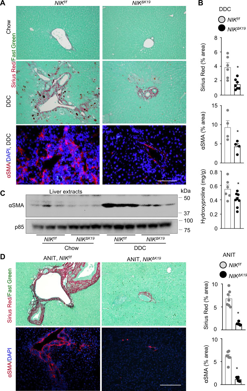
Fig. 5. Ablation of NIK in cholangiocytes protects against liver fibrosis.
A, B NIKΔK19 and NIKf/f male mice were fed a DDC diet for 4 weeks. Liver sections were stained with Sirius red/fast green or antibody to αSMA. A Representative images. Scale bar: 200 μm. B Sirius red (NIKf/f: n = 6 mice, NIKΔK19: n = 7 mice) and αSMA HSC (n = 4 mice per group) areas were measured and normalized to total areas. Liver hydroxyproline levels were measured and normalized to liver weight (NIKf/f: n = 6 mice, NIKΔK19: n = 8 mice). C Mice were fed a chow or DDC diet for 4 weeks. Liver extracts were immunoblotted with anti-αSMA and p85 (loading control) antibodies. Each lane represents one individual animal (n = 3 mice per group). D NIKΔK19 (n = 6 mice) and NIKf/f (n = 7 mice) male mice were fed an ANIT diet for 3 weeks. Liver sections were stained with Sirius red/fast green or anti-αSMA. Scale bar: 200 μm. Sirius red and αSMA areas were normalized to total areas. Data are presented as mean ± SEM. *p < 0.05, 2-tailed student’s t-test. Source data are provided as a Source Data file.

