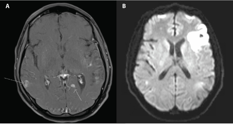
Figure 1. MRI and MRA findings of diffuse focal lesions and infarction.
A. Nodular foci of enhancement on MRI on T1-weighted image in the axial plane indicated by arrows concerning metastasis, infection, or vasculitis lesions. B. New area of infarct in the left frontal region on MRA on T2-weighted image in the axial plane. MRA: magnetic resonance angiogram.

