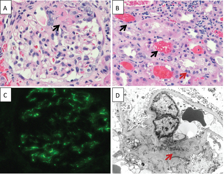
Figure 3. Kidney biopsy pathology.
A. Glomerulus with a small segment of fibrinoid tuft necrosis (black arrow) on light microscopy (HE stain, 200×). B. Several red blood cell casts (black arrows) located in tubular lumens and a tubule with marked cytoplasmic vacuolation of the tubular epithelial cells (red arrow) (HE, 200×). C. Mild (1+) granular staining for C3 in the mesangial areas of the glomerulus on immunofluorescence (200×). D. Electron microscopy image showing a mesangial region with very small, rare electron-dense deposits (red arrow). HE: hematoxylin and eosin stain.

