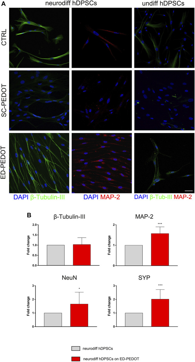
FIGURE 5.
Evaluation of neurogenic differentiation of hDPSCs on PEDOT: PSS films. The expression of the neuronal markers —Tubulin-III (green) and MAP-2 (red) in hDPSCs cultured in neurogenic medium for 7 days on both types of PEDOT:PSS films are shown by representative immunofluorescence images. Nuclei were counterstained with DAPI. hDPSCs differentiated on plastic culture plates were used as controls. Scale bar: 20 μm (A). Real-time PCR analysis showing fold increase of mRNA levels of —Tubulin-III, MAP-2, NeuN and Synaptophysin (SYP) in hDPSCs after 3 weeks of neurogenic induction on ED-PEDOT films. Data represent mean ± SD of fold change obtained from three independent experiments. * p < 0.05, *** p < 0.001 vs. neurodiff hDPSCs (i.e., on plastic culture plates) (B).

