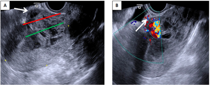
Fig. 2. (A) Transvaginal grayscale ultrasound demonstrates heterogeneous mass inside the uterine cavity protruding anteriorly through the scar tissue (arrow). Both the serosal line (red line) and uterine cavity line (green line) are crossed. (B) Colour Doppler imaging reveals intense vascularity in the vesicouterine space (arrow), involving posterior wall of urinary bladder.

