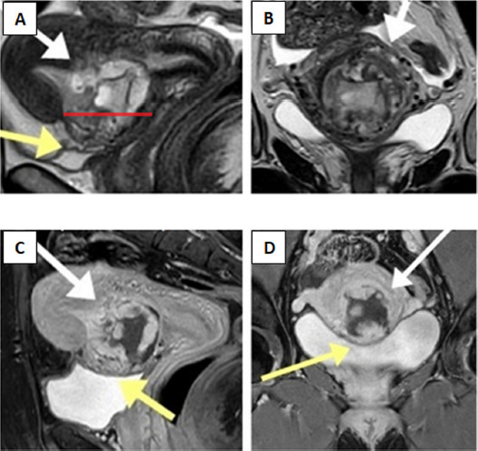
Fig. 3. (A) Sagittal T2-weighted magnetic resonance image demonstrates gestational sac as heterogeneous mass in the anterior part of lower uterine wall within the prior caesarean scar site, extending into the uterine cavity (white arrow). The scar margins are separated (red line) following infiltration by ectopic tissue. The “tenting sign’’ (yellow arrow) suggests trophoblast invasion into the wall of urinary bladder. (B) Coronal T2-weighted sequence demonstrates the disruption of thin myometrium secondary to pathological infiltration (white arrows). (C, D) Sagittal and coronal T1-weighted delayed post-contrast magnetic resonance images with full urinary bladder show no “tenting sign’’ (yellow arrows) evident in A image, therefore transmural trophoblast growth into the urinary bladder can be excluded.

