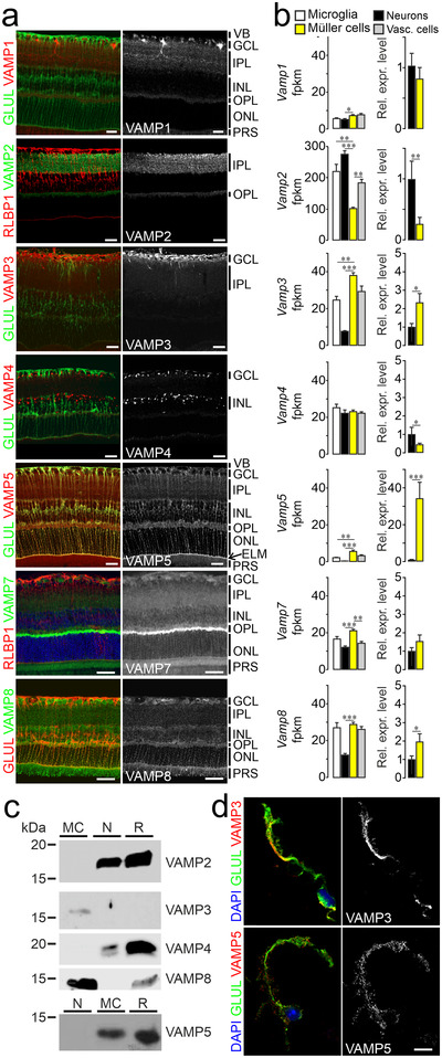
FIGURE 1.

Expression of VAMP3, 5 and 8 by retinal Müller cells (a) False‐colour and greyscale confocal micrographs of retinal sections from adult mice subjected to double‐immunohistochemical staining of the indicated VAMPs and of the Müller cell markers GLUL and RLBP1. Positions of selected ocular structures and retinal layers are indicated: VB, vitreous body; GCL, ganglion cell layer; IPL, inner plexiform layer; INL, inner nuclear layer; OPL, outer plexiform layer; ONL, outer nuclear layer; ELM, external limiting membrane (arrow); PRS, photoreceptor segments. Scale bars: 20 μm. (b) Left, average counts of indicated Vamp transcripts obtained by RNAseq from lysates of indicated types of acutely isolated, immunoaffinity‐purified retinal cells (n = 3–4 preparations; Mann‐Whitney test; whiskers indicate SEM). Fpkm, fragments per kilobase million. Right, mean relative expression levels of indicated Vamps in Müller cells compared to neurons. (c) Representative immunoblots showing the presence of selected VAMPs in indicated populations of acutely isolated immunoaffinity‐purified retinal cells (MC, Müller cells; N, neurons; R, retinal lysates). For each protein tested, equal amounts of total protein from indicated samples were loaded. (d) False‐colour and greyscale micrographs of acutely isolated immunoaffinity‐purified Müller cells subjected to nuclear (blue) and double‐immunocytochemical staining of VAMP3 and VAMP5 (red) and GLUL (green). Scale bar: 10 μm.
