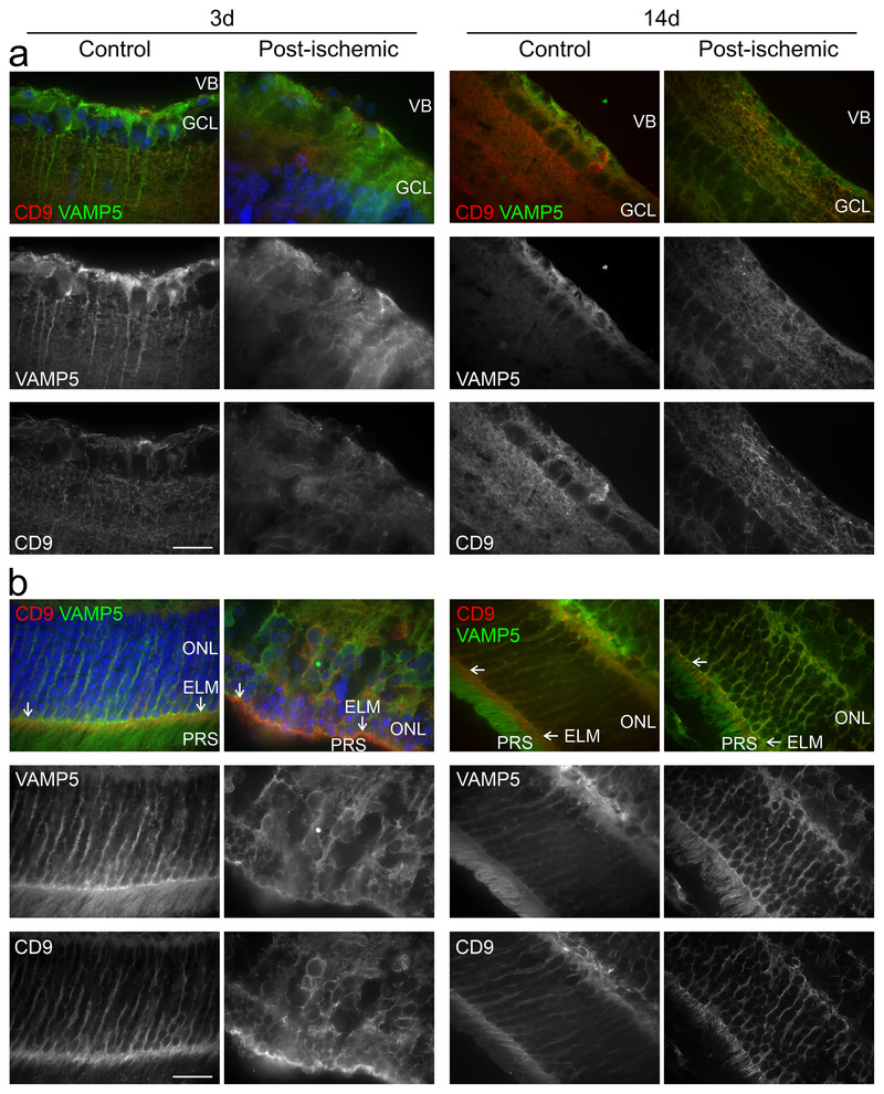
FIGURE 12.

Post‐ischemic changes in the retinal distribution of VAMP5 and CD9 False‐colour and corresponding greyscale confocal micrographs showing the inner (a) and outer (b) retinae from control eyes and from post‐ischemic eyes at 3 (left) and 14 (right) days after transient ischemia. Retinal sections were subjected to double‐immunohistochemical staining of VAMP5 (green) and of CD9 (red). VB, vitreous body; GCL, ganglion cell layer; ONL, outer nuclear layer; ELM, external limiting membrane (white arrows); PRS, photoreceptor segments. Scale bar: 40 μm.
