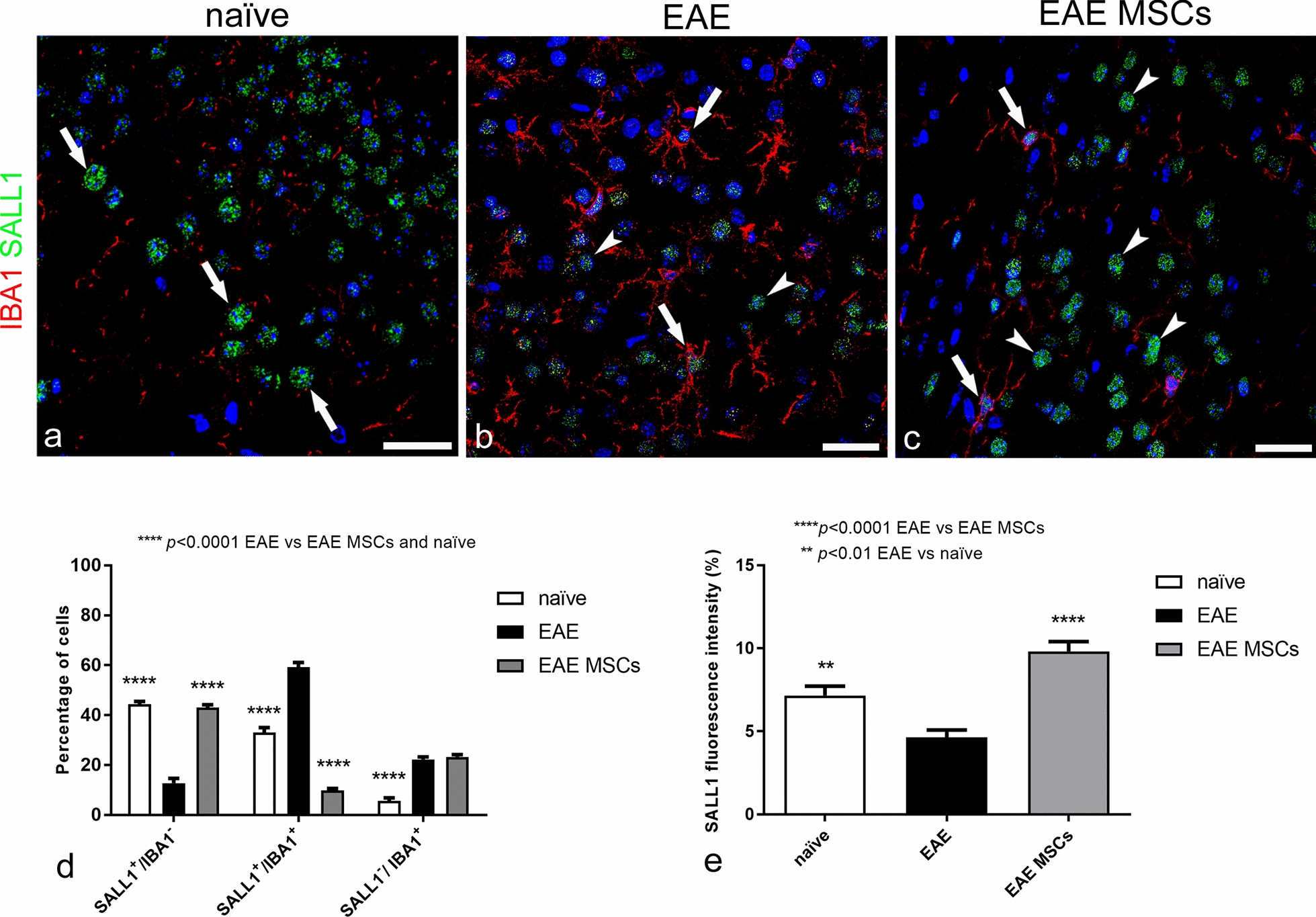
Fig. 6.

Representative images of neocortex sections from naïve (a), EAE-affected (b; cs 2.0) and EAE-affected MSC-treated (c; cs 1.5) mice, sacrificed 24 h after MSC treatment, double immunostained for IBA1 and SALL1; morphometric analyses of IBA1/SALL1 cell populations and SALL1 fluorescence intensity (d, e). a SALL1 specifically marks the nucleus of surveillant microglia, which express low levels of IBA1 (SALL1+/IBA1− cell population, arrows). b SALL1+/IBA1+ (arrows) and SALL1−/IBA1+ (arrowheads) cell populations are both recognizable. c Surveillant SALL1+/IBA1− microglia (arrows) prevail on SALL1+/IBA1+ reactive microglia (arrowhead). d In EAE-affected mice, the percentage of surveillant SALL1+/IBA1− microglia strongly declines in favor of a significant increase in reactive SALL1+/IBA1+ microglia and SALL1−/IBA1+ monocytes/macrophages. In EAE-affected MSC-treated mice, the percentage of SALL1+/IBA1− is similar to the level observed in naïve mice, whereas the increase in SALL1+/IBA1+ microglia observed in EAE-affected mice appears reverted by the treatment with MSCs; note the equal elevated percentage of SALL1−/IBA1+ monocytes/macrophages in both treated and not treated EAE-affected mice. e Fluorescence intensity for SALL1 is significantly higher in naïve mice microglia compared to EAE-affected mice; indeed, the highest levels are seen in EAE-affected MSC-treated mice. Data are reported as means ± SD (n = 3, n = 4, n = 4; 3 sections each), and the Bonferroni post-test was used to compare all groups after two-way ANOVA (d) or one-way ANOVA (e). Clinical score of EAE-affected mice (mean cs 2.1) and MSC-treated mice (mean cs 2.0). The TOPRO-3 nuclear counterstaining. TOPRO-3 nuclear counterstaining. Scale bars: a–c 25 µm
