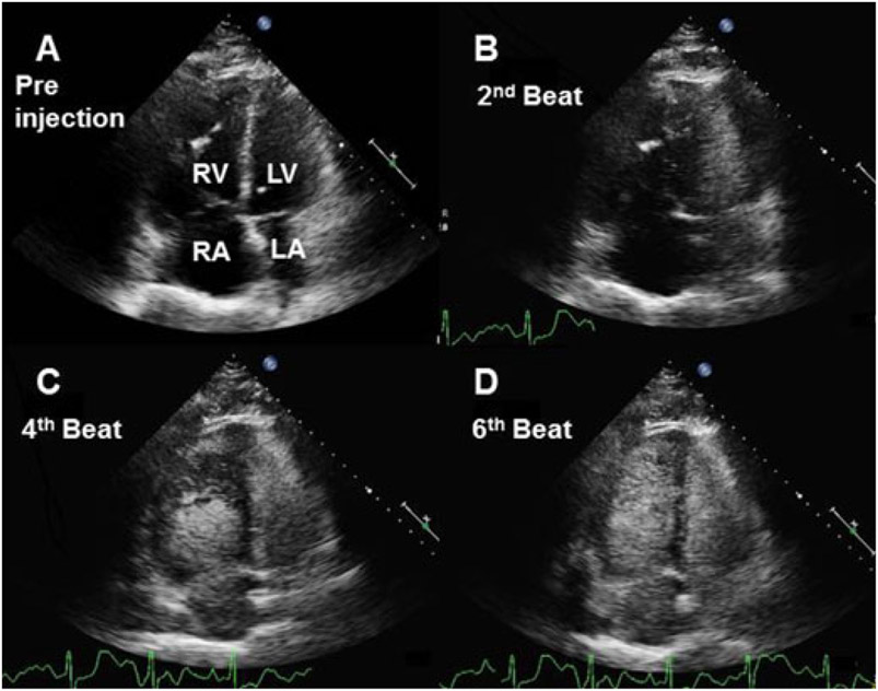
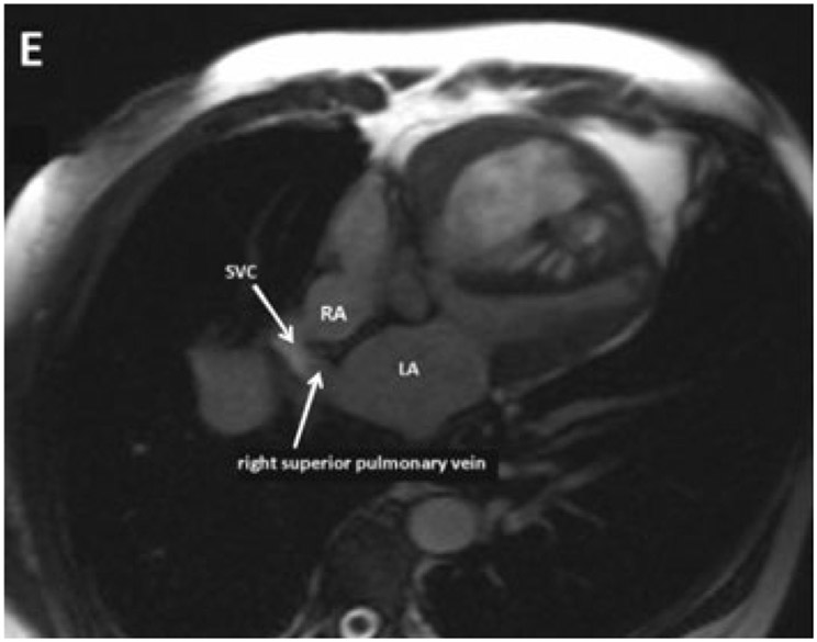
A 68-year-old man with a history of shortness of breath and hypoxia was undergoing workup for severe pulmonary hypertension. He was referred to pulmonary hypertension clinic for the first time. An echocardiography (iE33, Philips Medical Systems, Bothell, WA, USA) with agitated saline was performed. The calculated pulmonary artery pressure was 113 mmHg which was associated with right ventricular hypertrophy and dilatation. On contrast injection with agitated saline, the bubbles surprisingly filled the left heart rapidly before the right side (Fig. 1 and movie clips S1 and S2). Based on this bubble study, a diagnosis of Eisenmeger’s syndrome from a sinus venosus atrial septal defect was made and later confirmed on cardiac magnetic resonance imaging (Avanto, Siemens Medical Solutions, Erlangen, Germany) (Fig. 2).
Figure 1.
Echocardiography with agitated saline injection in a patient with a sinus venosus atrial septal defect. A. shows a four-chamber view prior to the injection of agitated saline, highlighting the right ventricular enlargement and hypertrophy associated with severe pulmonary hypertension. B.-D. show the progress of the bubbles after the 2nd, 4th, and 6th beats, respectively. LA = left atrium; LV = left ventricle; RA = right atrium; RV = right ventricle.
Figure 2.
The MRI of the sinus venosus atrial septal defect. LA = left atrium; RA = right atrium; SVC = superior vena cava.
The fact that the bubbles first appear on the left side suggests that the shunt occurs above the level of the atria. Because of high right-sided pressures in this case, flow from the superior vena cava (SVC) preferentially travels through the defect to the right superior pulmonary vein and left atrium.
A residual left SVC with a direct connection to the left atrium via an unroofed coronary sinus was also considered as it would have also shown a similar filling pattern with agitated saline. This defect was excluded, however, because the agitated saline was serendipitously injected into the right hand.
In this case, the shunt was not diagnosed until late in life. He had been treated for years with vasodilators for presumed primary pulmonary hypertension. Unfortunately, he died 3 months later from severe Eisenmeger’s syndrome. In this case, the initial echocardiography with agitated saline was essential in making the diagnosis, but because of the large right-to-left shunt, further bubble studies should be avoided in patients with known Eisenmeger’s syndrome.
Supplementary Material
Movie clip S1. The first injection of agitated saline in a patient with sinus venosus atrial septal defect and Eisenmenger’s syndrome.
Movie clip S2 The second injection of agitated saline in a patient with sinus venosus atrial septal defect and Eisenmenger’s syndrome. Note a few residual bubbles in the right atrium at the beginning of the study are left over from the first injection.
Footnotes
Supporting Information
Additional Supporting Information may be found in the online version of this article:
Associated Data
This section collects any data citations, data availability statements, or supplementary materials included in this article.
Supplementary Materials
Movie clip S1. The first injection of agitated saline in a patient with sinus venosus atrial septal defect and Eisenmenger’s syndrome.
Movie clip S2 The second injection of agitated saline in a patient with sinus venosus atrial septal defect and Eisenmenger’s syndrome. Note a few residual bubbles in the right atrium at the beginning of the study are left over from the first injection.


