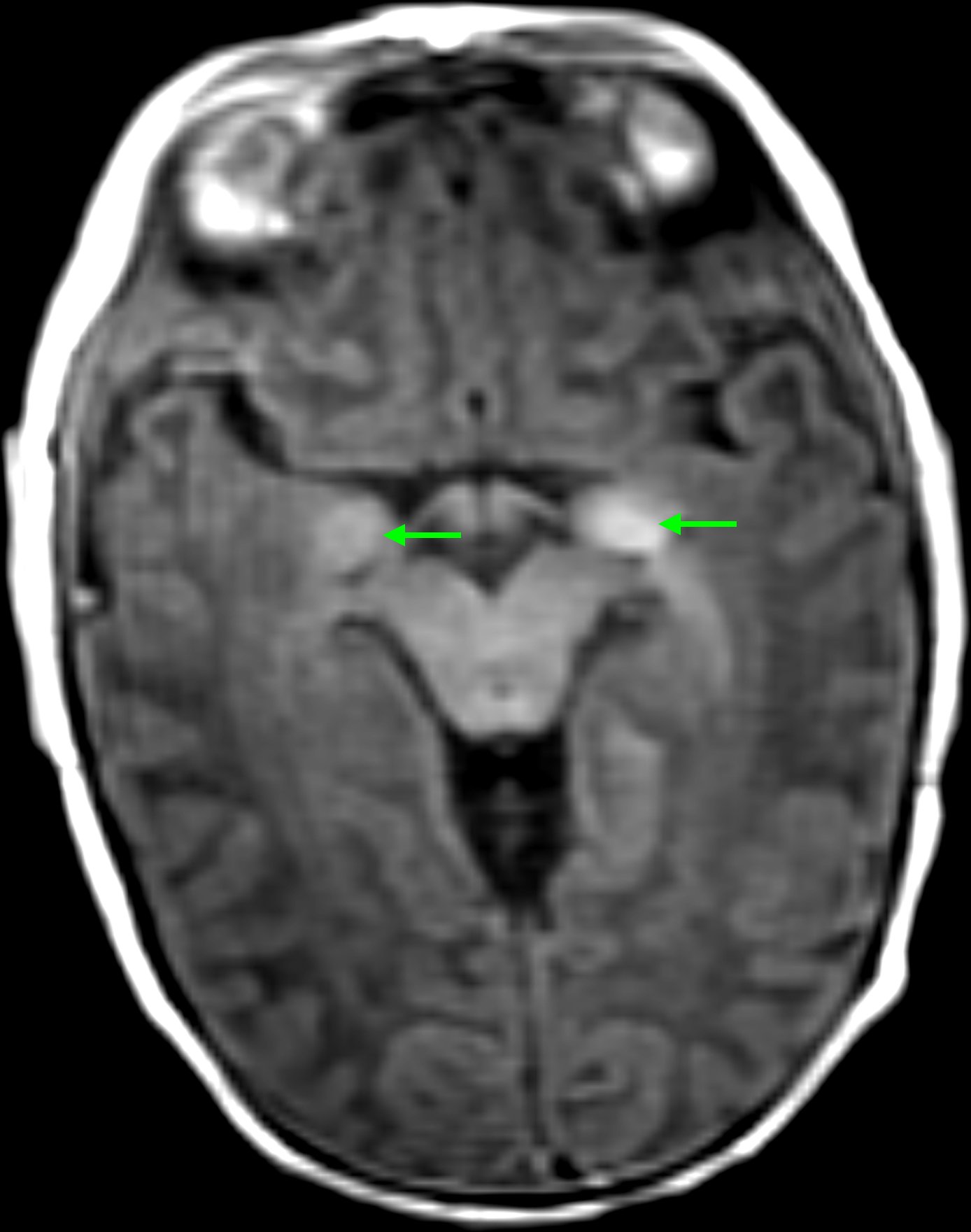
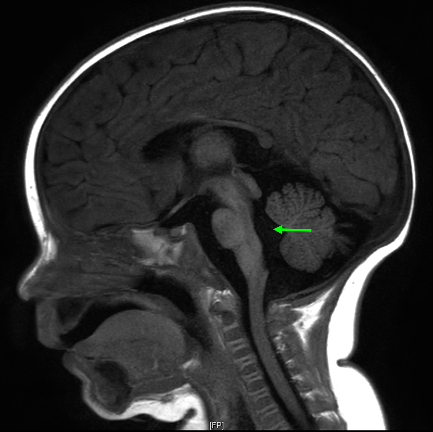
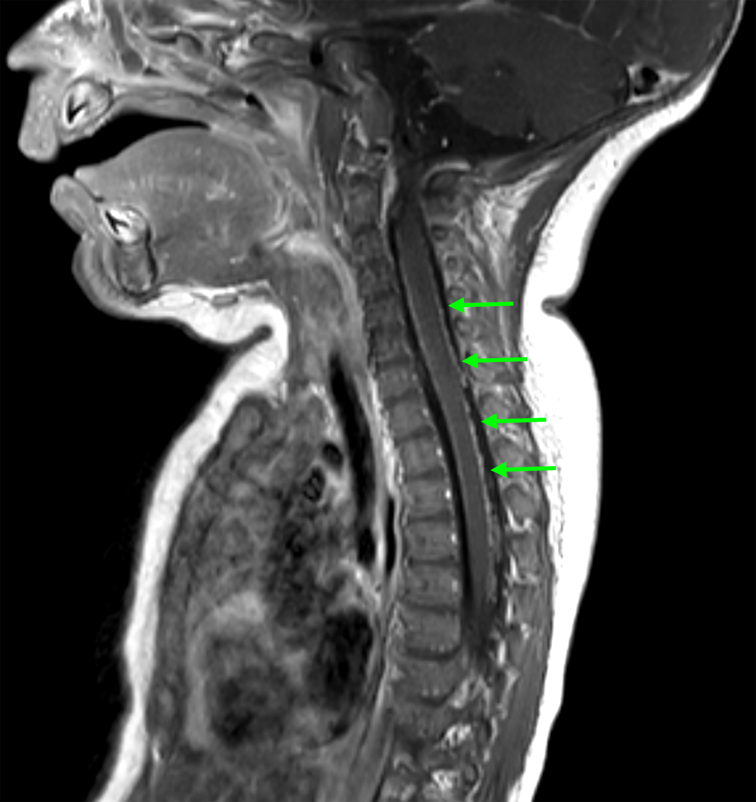
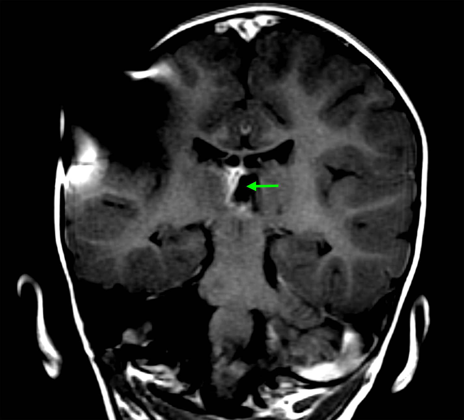
Figure 1:


(A) Axial T1 MRI brain obtained shortly after birth, demonstrating T1 shortening in the cerebellum and brainstem (arrows), appearance radiographically consistent with NCM; (B) Sagittal T1 MRI brain at five months of age demonstrating hydrocephalus. Enlargement of the fourth ventricle is evident (arrow); (C) Sagittal T1 post-contrast MRI of the cervical and upper thoracic spine, obtained at the time patient presented with symptomatic hydrocephalus. Diffuse leptomeningeal enhancement is evident along the ventral and dorsal surfaces of the spinal cord (arrows).
