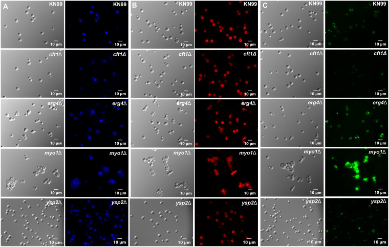
FIG 6.
Deletion of MYO1 altered yeast cell morphology, and deletions of either ERG4 or MYO1 increased WGA staining. Cell wall staining phenotypes of KN99 and the cft1Δ, erg4Δ, myo1Δ, and ysp2Δ deletion strains. Cells were incubated with calcofluor white (A), cibacron red (B), or wheat germ agglutinin-Alexa Fluor 488 (C). A differential interference contrast (DIC) image of the same slide section is shown to the left of each fluorescence image.

