FIG 7.
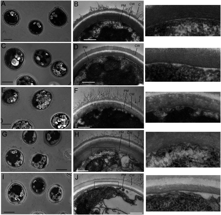
The cell walls of all deletion strain cells are intact. Transmission electron microscopy of KN99 cells at a magnification of ×3,000 (A), KN99 cells at ×25,000 (B), cft1Δ cells at ×3,000 (C), cft1Δ cells at ×25,000 (D), erg4Δ cells at ×3,000 (E), erg4Δ cells at ×25,000 (F), myo1Δ cells at ×3,000 (G), myo1Δ cells at ×25,000 (H), ysp2Δ cells at ×3,000 (I), and ysp2Δ cells at ×25,000 (J). The boxed areas in the ×25,000 magnification images are shown enlarged to the right of the original images. The scale bars show 2 μm at ×3,000 magnification and 500 nm at ×25,000 magnification. PM, plasma membrane; CW, cell wall; C, capsular material. Cells were grown overnight in YPD at 30°C prior to preparation for imaging. Sections were stained with uranyl acetate and lead acetate.

