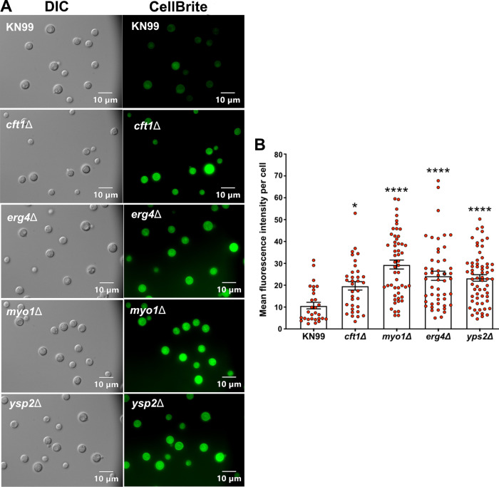
FIG 8.
The membranes of the deletion strains were more exposed to membrane-specific dyes than the membranes of wild-type cells. Overnight cultures of KN99 and the four deletion strains were stained with CellBrite fix 488. The stained cells were directly imaged without fixing. (A) Representative DIC and fluorescent images of the five strains. (B) Mean fluorescence intensity was based on measuring the fluorescence of 30 or more cells per strain. The differences in mean fluorescence were evaluated using one-way ANOVA with Dunnett’s post hoc test. Error bars show standard deviations. *, P < 0.05; ****, P < 0.001.

