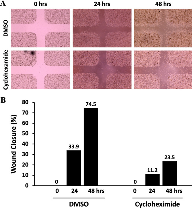
FIG 2.

Analysis of HeLa cell migration in the in vitro wound healing assay. (A) Micrograph of wound healing assays with DMSO and cycloheximide. Assays were performed with an ibidi culture insert in 4-well 35-mm dishes. (Top) Microscopy images of wound closure of DMSO-treated control cells at 0, 24, and 48 h after culture insert removal. (Bottom) Images were also taken at 0, 24, and 48 h after cycloheximide treatment. Cycloheximide-treated cells showed less migration than DMSO-treated counterparts. (B) Percentage of wound closure at 0, 24, and 48 h postinjury. The center wound areas were selected from micrographs, and wound closure was quantified using ImageJ software.
