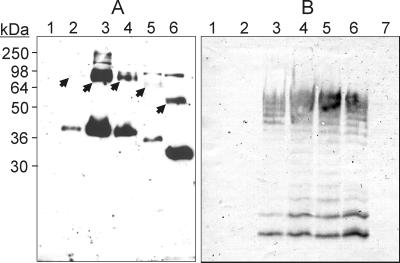
FIG. 3.
(A) Detection of WecA derivatives tagged with the FLAG epitope by immunoblotting with MAb M2. JM109DE3 cell lysates were separated by SDS-PAGE, transferred to nitrocellulose membranes, and processed as described in the text. Lanes: 1, pAA8; 2, pAA11; 3, pAA12; 4, pAA14; 5, pAA15; 6, pAA16. Arrows indicate polypeptides with higher molecular masses (barely visible in lanes 2 and 5) that may represent oligomers of WecA. The positions of the following molecular mass standards are shown: myosin (250 kDa), bovine serum albumin (98 kDa), glutamic acid dehydrogenase (64 kDa), alcohol dehydrogenase (50 kDa), carbonic anhydrase (36 kDa), and myoglobin (30 kDa). (B) Immunoblot of LPS samples prepared from MV501 (lanes 1 to 5 and 7) and VW187 (lane 6), which were reacted with anti-O7 antibodies. MV501 was transformed with the various wecA derivatives as follows: lane 1, pAA16; lane 2, pAA15; lane 3, pAA14; lane 4, pAA12; lane 5, pAA11.

