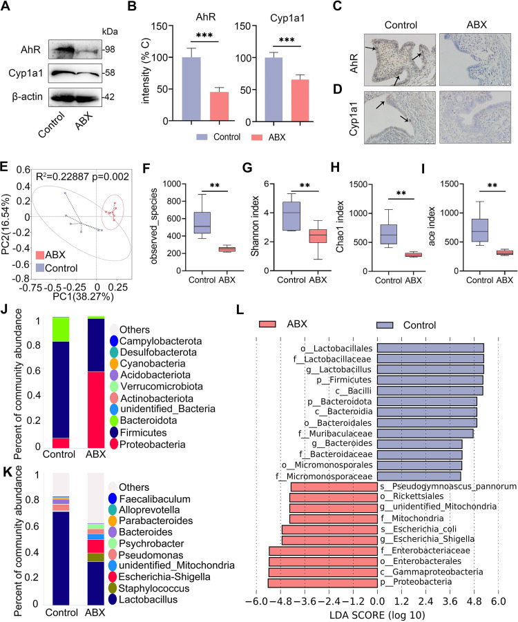
FIG 3.
Gut dysbiosis impairs uterine AhR activation and reduces the abundance of intestinal AhR producers in mice. The mice were treated with ABX (1 g/L ampicillin, metronidazole, and neomycin sulfate and 0.5 g/L vancomycin) for 3 weeks, and fecal and uterine samples were harvested for determination. (A) Levels of uterine AhR and Cyp1a1 in control and ABX-treated mice were assessed by Western blotting. (B) Intensities of AhR and Cyp1a1 in the indicated mice were determined (n = 5). (C) Representative images of AhR antibody-stained uterine sections from control and ABX treated mice are shown. The arrow indicates positively stained cells (scale bar = 20 μm). (D) Representative images of Cyp1a1 antibody-stained sections are shown. The arrow indicates positively stained cells (scale bar = 20 μm). (E) Principal coordinates analysis (R2 = 0.22887, P = 0.002) shows distinct gut microbial structure between the control and ABX treatment groups based on unweighted UniFrac distances (n = 6). (F-I) Alpha diversity analyses, including observed species (F), Shannon (G), Chao1 (H), and ace index (I), were performed from different treatment groups (n = 6). (J-K) Gut bacterial compositions at the phylum (J) and genus (K) levels are shown. (L) Linear discriminant analysis (LDA) effect size (LEfSe) was performed to show the most differentially significant bacterial taxa enriched in the control and ABX groups (log10 LDA score > 4). Data are expressed as the mean ± SD (B) or boxplot (F-I), and two-tailed Student's t test (B) and Mann-Whitney U test were performed for statistical analysis (F-I). **, P < 0.01; and ***, P < 0.001 indicate significant differences. ABX, cocktail of antibiotics.

