
FIG 4.
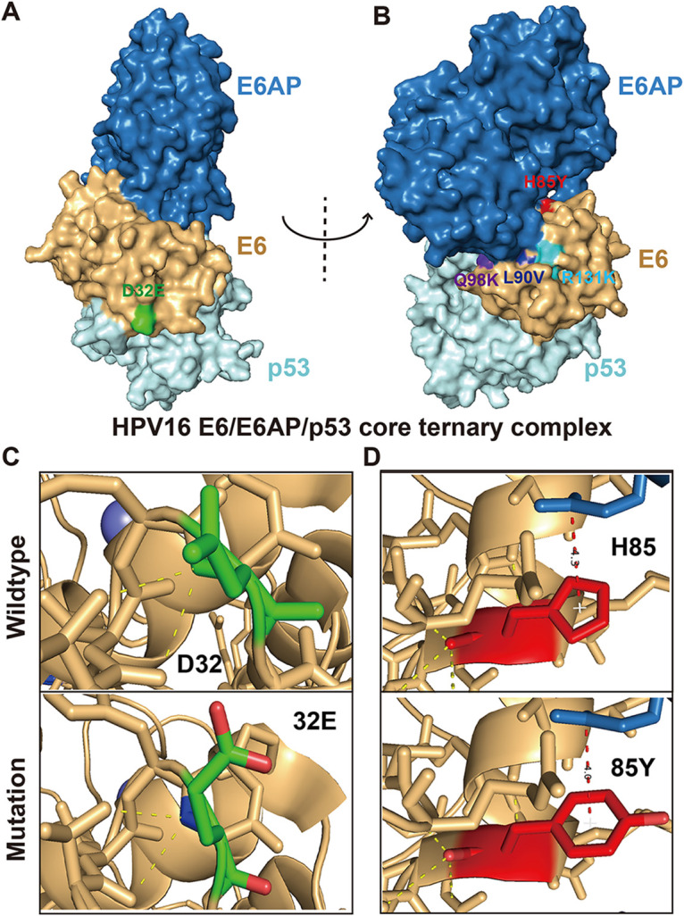
The 3D structural analysis of HPV16 E6 mutations. (A, B) Structure of the HPV16 E6/E6AP/p53 core ternary complex (25) (blue, E6AP peptide; brown, HPV16 E6; light cyan, p53 core). The mutations are marked on the surface by different colors (green, D32E; red, H85Y; dark blue, L90V; purple, Q98K; powder blue, R131K). (C) The 3D prediction structures of E6–p53 interface associated with D32E mutation. (D) The 3D prediction structures of E6–E6AP interface associated with H85Y mutation.