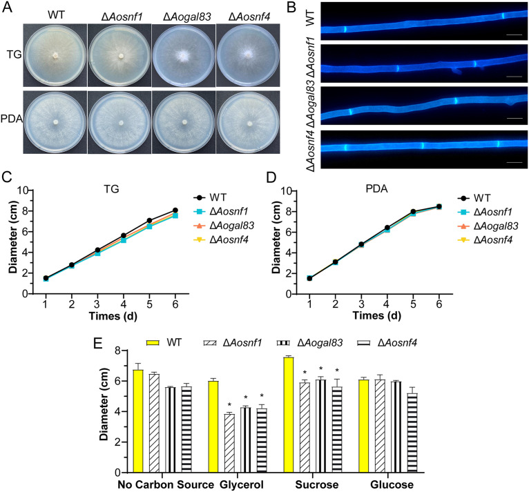
FIG 2.
Colony morphology and diameters between the wild-type (WT) and AMPK complex mutant (ΔAosnf1, ΔAogal83, and ΔAosnf4) strains. (A) Colonies of strains cultured on TG and PDA plates for 6 days at 28°C. (B) Mycelia of the WT and mutant strains on PDA. Mycelia were stained with calcofluor white (CFW). Scale bar, 5 μm. (C) Colony diameters of the WT and mutant strains cultured on TG plates for 6 days. (D) Mycelia of the WT and mutant strains on PDA. (E) Colony diameters of the WT and mutant strains cultured on CDA medium with different carbon sources for 6 days. *, significant difference between the mutant and WT strains (P < 0.05).

