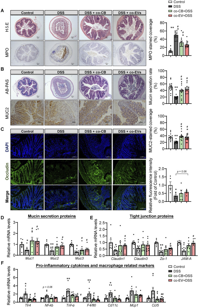
FIG 7.
C. butyricum-derived extracellular vesicles administration attenuated the colonic barrier damage. (A) H&E staining of the colon with summarized histological score and immunohistochemical analysis of MPO with summarized MPO stained rate (n = 5). (B) Alican blue-PAS staining of the colon with its mucin secretion rate and immunohistochemical analysis of MUC2 with MUC2-stained coverage (n = 5). Scale bar: 200 μm. (C) Immunofluorescence staining of tight junction protein Occludin expression in the colonic section from mice (n = 5). Blue: DAPI; Green: Occludin. Scale bar: 100 μm. (D) Relative mRNA levels of Mucin secretion proteins, tight junction proteins, and proinflammatory related gene expression (n = 7 to 8). *, P ≤ 0.05; **, P ≤ 0.01 versus the control group, #, P ≤ 0.05; ##, P ≤ 0.01 versus the DSS group.

