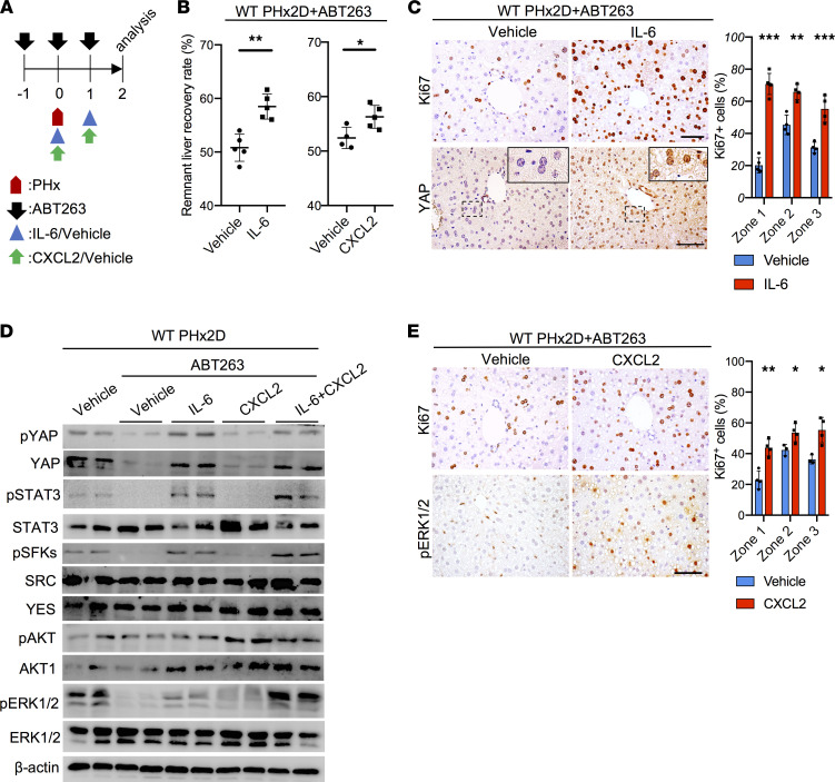
Figure 4. IL-6 or CXCL2 promotes liver regeneration in ABT263-treated mice.
(A) Experimental scheme of ABT263-treated WT male mice subjected to PHx with vehicle (PBS), IL-6 (50 μg/kg/d), or CXCL2 (15 μg/kg/d) injections. (B) The remnant liver recovery rates of the indicated livers were measured 2 days after PHx (PHx2D; n = 4–5). (C) Liver sections (zone 1) of WT mice subjected to ABT263 and PHx followed by injections of vehicle or IL-6 were stained with anti-Ki67 or anti-YAP antibodies 2 days after PHx. The percentages of Ki67+ hepatocytes in all 3 zones were quantified by cell counting (n = 4). (D) Immunoblots of protein extracts from whole liver lysates of indicated mice were probed with the indicated antibodies. ABT263-treated mice were injected with vehicle, IL-6, CXCL2, or both on days 0 and 1 after PHx, then harvested on day 2 for analysis. (E) Liver sections of WT mice subjected to ABT263 and PHx followed by injections of vehicle or CXCL2 were stained with anti-Ki67 (zone 1) or anti–p-ERK1/2 antibodies 2 days after PHx. The percentages of Ki67+ hepatocytes in all 3 zones were quantified by cell counting (n = 4). Scale bar: 50 μm. Data are expressed as mean ± SD. *P < 0.033, **P < 0.002, ***P < 0.001, Student’s t test.

