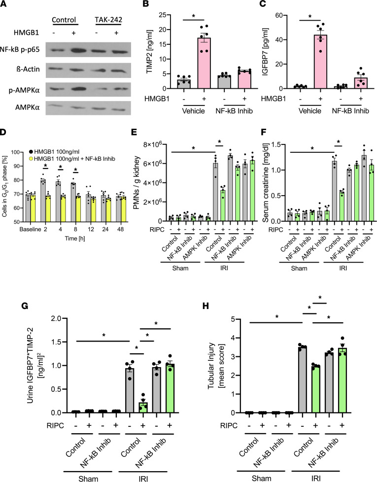
Figure 6. HMGB1 induces NF-κB and AMPKα activation.
(A) Isolated murine renal tubular epithelial cells were incubated with HMBG1 (0.1 μg/mL) or HMGB1 plus TLR4 inhibitor (TAK-242). Activation of NF-κB and AMPKα was detected by Western blotting for NF-κB p-p65 and β-actin as loading control, as well as p-AMPKα and total AMPKα (exemplary blots). Isolated murine renal tubular epithelial cells were incubated with HMBG1 (0.1 μg/mL) or HMGB1 plus NF-κB inhibitor (Bay-117082). (B and C) TIMP-2 and IGFBP7 were analyzed by ELISAs (n = 6). (D) Isolated murine renal tubular epithelial cells were treated with HMBG1 (0.1 μg/mL) or HMGB1 in combination with NF-κB inhibitor (Bay-117082). The proportion of cells in G0/G1 phase was analyzed by measuring cellular DNA content by flow cytometry (n = 6). After induction of general anesthesia WT mice received either 3 cycles RIPC or control procedure. Some mice received a NF-κB inhibitor (Bay-117082, 10 mg/kg i.p.) or AMPKα inhibitor before RIPC. Twenty-four hours after IRI induction, mice were sacrificed. (E) The recruitment of neutrophils (PMNs) into the kidney was analyzed by flow cytometry (n = 4). (F) Serum creatinine levels were measured by a photometric assay (n = 4). (G) The biomarkers TIMP-2 and IGFBP7 were measured in urine samples 24 hours after inducing renal IRI. (H) Renal tubular injury score was assessed based on histology (n = 4). One-way ANOVA followed by Bonferroni testing was used for statistical analysis; *P < 0.05.

