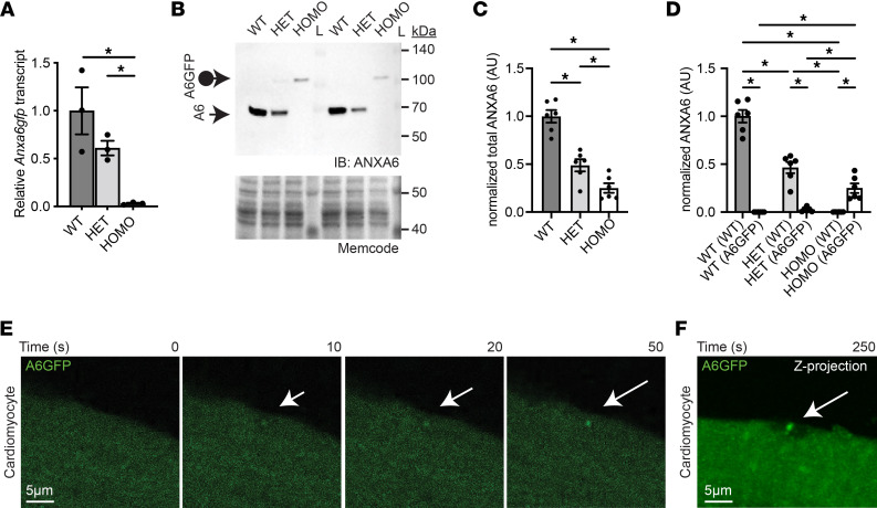
Figure 3. Genomically encoded annexin A6GFP localizes at the site of cardiomyocyte membrane injury.
(A) Quantitative PCR demonstrates reduced Anxa6 levels in heart lysates from heterozygous and homozygous Anxa6gfp mice compared with WT controls. (B–D) Anti–annexin A6 immunoblots demonstrate reduced ANXA6 protein levels in cardiac ventricle lysates from heterozygous and homozygous Anxa6gfp mice. The loading control is a 42 kDa band detected by MemCode reversible protein stain. (E) Adult ventricular cardiomyocytes were isolated from homozygous Anxa6gfp mice and subsequently laser-damaged. annexin A6GFP (shown in green) quickly localizes to the cardiomyocyte repair cap (white arrow). (F) Z-projection of a homozygous Anxa6gfp cardiomyocyte illustrating annexin A6GFP repair cap (white arrow) above the annexin-free zone at the site of injury 250 seconds after cardiomyocyte wounding. Scale bar: 5 μm. n = 6 mice per genotype. n > 12 cells from 5 isolations. *P < 0.05 by 1-way ANOVA.

