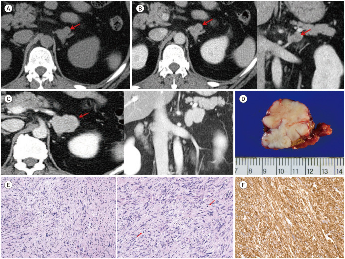
Fig. 1. Primary adrenal leiomyosarcoma in a 48-year-old woman.
A, B. CT images reveal a heterogeneously enhancing nodule (arrows) in the left adrenal gland. Pre-enhanced axial section (A), post-enhanced axial and coronal sections (B).
C. Abdominal CT images after two years. The CT images (axial sections and coronal sections) demonstrate an increase in the size of the homogeneously enhancing lobulated mass (arrow) in the left adrenal gland.
D. Photograph of the gross pathologic specimen. A well-defined, unencapsulated, and lobulated tumor, measuring 4.5 × 3 × 3 cm, is noted. The cut surface is white, firm, and fibrotic.
E, F. Photomicrograph images of adrenal leiomyosarcoma. The tumor is composed of intersecting fascicles of spindle cells. The tumor cells show pleomorphic nuclei with mitotic figures (arrows). Some of the tumor cells show elongated nuclei (hematoxylin and eosin × 100 and magnification × 200) (E). Immunohistochemical staining for smooth muscle actin is positive (× 200). Pathologic diagnosis was adrenal leiomyosarcoma (F).

