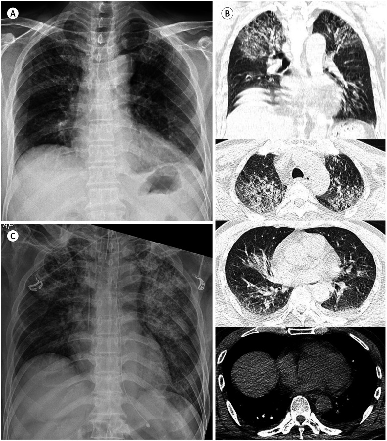
Fig. 1. A 57-year-old man with COVID-19 pneumonia.
A. Chest radiograph taken just before the transfer to our institution shows subtle GGOs in both upper lung zones and right middle lung zone.
B. Axial and coronal non-enhanced chest CT images, taken immediately after A, show large areas of mixed GGO and consolidation involving both upper lungs with underlying pulmonary emphysema. A small amount of bilateral pleural effusions and pericardial effusion is also visible.
C. Follow-up chest radiograph taken after the patient's arrival at our institution (12 hours after A and B) shows a marked increase in the extent and density of GGOs, progressed into diffuse consolidation in both lungs. Endotracheal intubation, central line insertion, and L-tube insertion were performed for further treatment.
GGO = ground glass opacity

