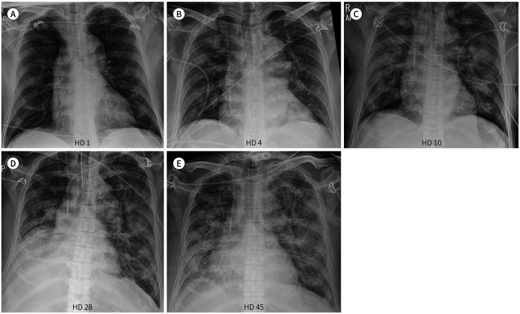
Fig. 3. A 61-year-old man with COVID-19 pneumonia.
A. Initial chest radiograph shows no significant abnormalities except cardiomegaly.
B. Chest radiograph on HD 4 shows new subtle patchy areas of GGO in the right upper and lower lung zones. Central line insertion was performed for further treatment.
C. Chest radiograph on HD 10 shows diffuse consolidation in both lungs, suggesting progression of pneumonia. Endotracheal intubation and L-tube insertion were performed for further treatment.
D. Chest radiograph on HD 28 shows no significant interval changes of diffuse consolidation in both lungs. Combined atelectasis of right middle and lower lobes is newly noted.
E. Chest radiograph on HD 45 shows an increase in extent of diffuse consolidation in both lungs, while combined atelectasis of right middle and lower lobes improves.
GGO = ground glass opacity, HD = hospital day

