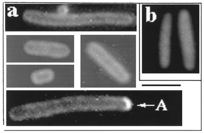
FIG. 2.
Distribution of Gfp-MinD in the absence of other Min proteins. Strain PB114/pSLR22 (ΔminCDE/Para-gfp::minD) was grown in the presence of 0.005% arabinose for 4 h at 30°C prior to fluorescence microscopy. (a) Fixed cells showing the peripheral pattern of Gfp-MinD fluorescence. A, polar arc. (b) Fixed cells; pSLR22 was replaced by pSLR21(Para-gfp).

