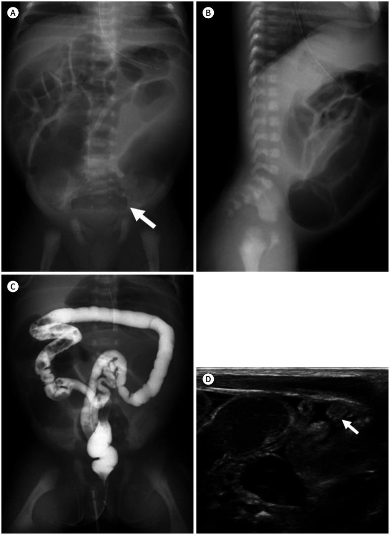
Fig. 11. A 2-day-old male neonate with ileal atresia.
A. On a simple radiograph, distended small bowel loops show the “multiple bubble sign.” A bulbous dilated segment (arrow) is noted at the lower abdomen.
B. In the lateral view, rectal gas is not noted. At this point, Hirschsprung's disease is hard to differentiate.
C. During enema, by using a water-soluble contrast material, decreased diameter of the colon loop is noted, which suggests an unused microcolon.
D. Dilated fluid-filled small bowel loops with a collapsed unused microcolon (arrow) can also be noted on ultrasonography.

