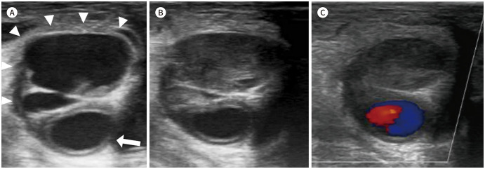
Fig. 2. A 67-year-old male with a right common femoral artery pseudoaneurysm.
A. The gray-scale ultrasound image shows a multiloculated pseudoaneurysm (arrowheads) from the right common femoral artery (arrow).
B, C. Ultrasound images show a pseudoaneurysm after the ultrasound-guided percutaneous thrombin injection. Complete thrombosis of the pseudoaneurysmal sac is achieved (B). There is no internal blood flow on color Doppler ultrasonography (C).

