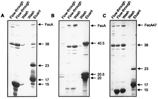
FIG. 3.
Binding of FecA to (His)10-FecR (A) and FecR-(His)6 (B) on a Ni-NTA agarose column and of FecAΔ47 to His10-FecR (C) on a Ni-NTA agarose column. The eluted fractions were subjected to SDS-PAGE (with 15% acrylamide gels) and stained with Coomassie brilliant blue. FecA, FecAΔ47 (designated FecA47), FecR-(His)6, (His)10-FecR, and their proteolytic cleavage products are indicated by arrows. Numbers indicate molecular masses in kilodaltons.

