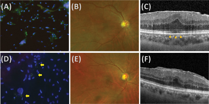
Fig 2.
Representative images of stained epiretinal membrane (ERM) specimens and retinal images from patients with primary ERM with (A-C) or without (D-F) ellipsoid zone (EZ) defects. (A) Epiretinal membrane specimen from a 64-year-old female patient with primary ERM with EZ defect, showing higher positive staining for sphingosine-1-phosphate (S1P). (B) Ultrawide field retinal photograph that shows no specific underlying retinal disease. (C) Image of spectral-domain optical coherence tomography (SD-OCT) that shows ERM with EZ defect (arrowheads). (D) Epiretinal membrane specimen from a 73-year-old female patient with primary ERM without EZ defect, showing lower positive staining for S1P. Only a couple of cells were stained with S1P (arrows). (E) Ultrawide field retinal photography that shows no specific underlying retinal disease. (F) Image of SD-OCT that shows ERM without EZ defect.

