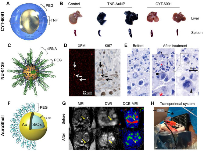
Fig. 2.
Representative therapeutic gold nanoconstructs in clinical trials. A Schematic representation of CYT-6091. B Comparison of ex vivo organ accumulation of non-PEGylated (TNF-AuNP) and PEGylated CYT-6091. Adapted with permission of ref 22. Copyright 2012 CytImmune. C Schematic representation of NU-0129. D Au elemental map of glioblastoma tumor sample and matching Ki67 staining after NU-0129 treatment. Arrows highlight gold accumulation regions within perivascular Ki67-positive cancerous cells. E Silver staining of glioblastoma tumors before and after NU-0129 treatment. Adapted with permission of ref 36. Copyright 2020 American Association for the Advancement of Science. F Schematic representation of AuroShell. Adapted with permission of ref 38. Copyright 2015 Elsevier. G Pre- and post-treatment images of patient with focal prostate cancer treated with AuroShell and photoablation. Imaging techniques: t2-weighted magnetic resonance imaging (MRI), diffusion-weighted imaging (DWI) and dynamic contrast-enhanced magnetic resonance (DCE-MRI) imaging. Arrows highlight tumoral region. H Picture of lasers introducers in thermocouple through transperineal grid. Adapted with permission of ref 40. Copyright 2019 National Academy of Science

