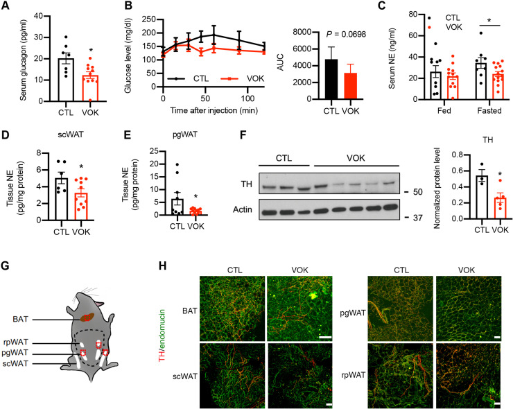
Fig. 4. Mice with SF1 neuron-specific OGT deletion show reduced activity of the sympathetic nervous system and impaired sympathetic innervation of scWAT.
(A) Serum glucagon levels of CTL and VOK mice after 15 hours of fasting. (B) Blood glucose levels of CTL and VOK mice after 2DG challenge. Quantification is shown on the right. (C) Serum norepinephrine levels of CTL and VOK mice fed ad libitum and after 15 hours of fasting. (D and E) Tissue norepinephrine levels in scWAT (D) and pgWAT (E) of CTL and VOK mice after 6 hours of fasting. (F) Western blot of tyrosine hydroxylase (TH) in scWAT of CTL and VOK mice after 6 hours of fasting. Densitometric quantification is shown on the right. (G) Workflow showing the process of tissue immunostaining of TH in adipose tissues of CTL and VOK mice at 7 months of age. Red dotted squares indicate the 2-mm3 tissue cubes used for immunostaining. (H) Representative immunostaining of TH (red) and endomucin (green) in BAT, scWAT, pgWAT, and rpWAT of CTL and VOK mice fed ad libitum. Scale bars, 100 μm. CTL: n = 7 to 9 and VOK: n = 10 to 15 for glucagon and norepinephrine enzyme-linked immunosorbent assay. CTL: n = 4 and VOK: n = 5 for the 2DG challenge. CTL: n = 3 and VOK: n = 3 for tissue immunostaining. Mice were fed a normal chow diet. The experiments were performed on CTL and VOK mice aging 14 to 20 weeks before body weight divergence. Data are shown as means ± SEM. *P < 0.05 by unpaired Student’s t test.

