. 2022 Aug 12;11:e77825. doi: 10.7554/eLife.77825

Figure 1. Screening of Nme1Cas9 orthologs activities through a GFP- activation assay.

(A) Schematic of the GFP-activation assay. A protospacer flanked by an 8 bp random sequence is inserted between the ATG start codon and GFP-coding sequence, resulting in a frameshift mutation. The library DNA is stably integrated into HEK293T cells via lentivirus infection. Genome editing can lead to in-frame mutation. The protospacer sequence is shown below. (B) The procedure of the GFP-activation assay. Cas9 and sgRNA expression plasmids were co-transfected into the reporter cells. GFP-positive cells could be observed if the protospacer is edited. (C) Twenty-five out of 29 Nme1Cas9 orthologs could induce GFP expression. The percentage of GFP-positive cells is shown. Reporter cells without Cas9 transfection are used as a negative control. Scale bar: 250 μm.

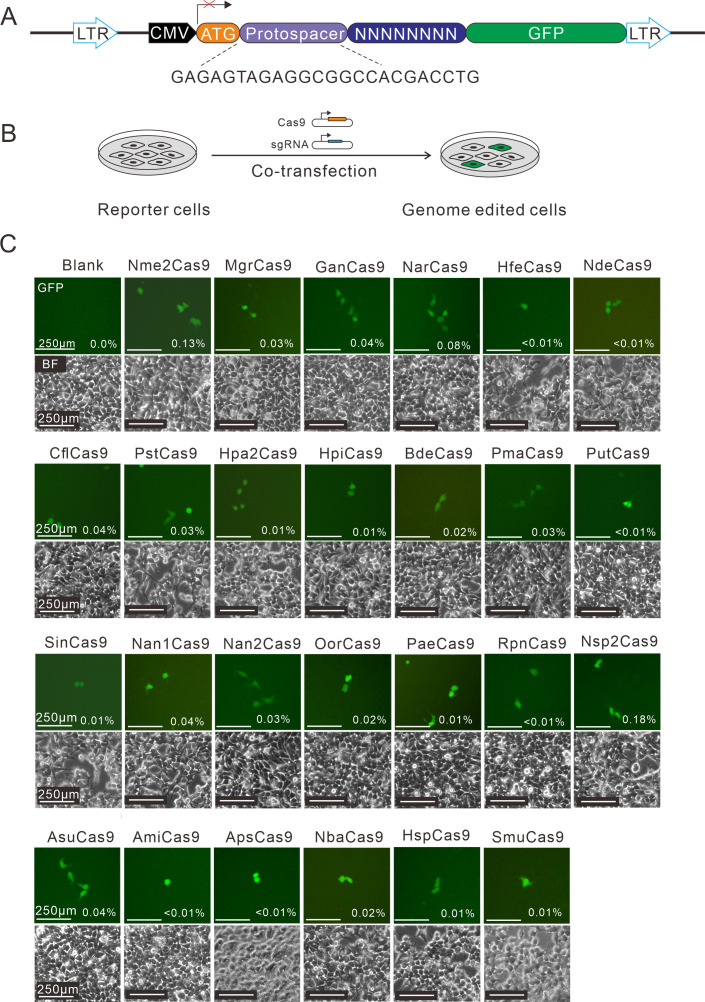
Figure 1—source data 1. The maps of all plasmids used in the study for Figure 1.

Figure 1.

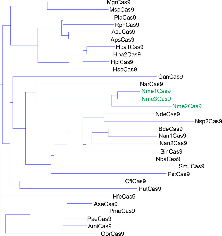
Figure 1—figure supplement 1. Phylogenetic tree of the selected Nme1Cas9 orthologs.

Figure 1—figure supplement 1.

The amino acid sequences of 29 selected Nme1Cas9 orthologs were aligned by Vector NTI. Nme1Cas9, Nme2Cas9, and Nme3Cas9 were used as reference and shown in green.
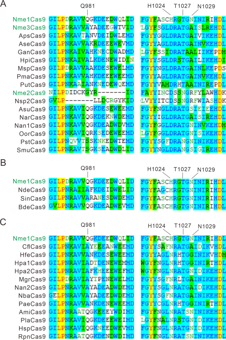
Figure 1—figure supplement 2. Alignment of the PI domain of Nme1Cas9 orthologs.

Figure 1—figure supplement 2.

The Nme1Cas9 orthologs contained aspartate (A), histidine (B), or asparagine (C) residues corresponding to the Nme1Cas9 H1024. The PI domains were aligned by Vector NTI. Amino acids crucial for PAM recognition are shown above. Nme1Cas9, Nme2Cas9, and Nme3Cas9 were used as reference and shown in green.
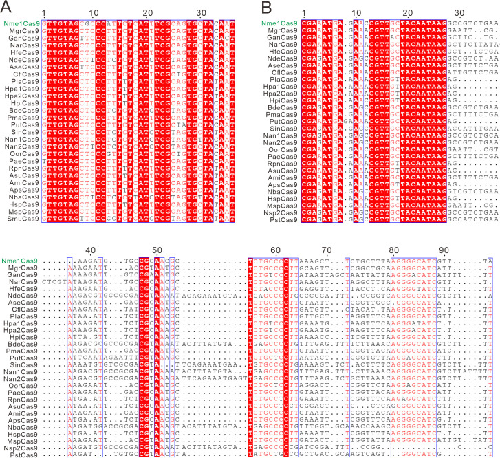
Figure 1—figure supplement 3. The alignment of direct repeats and tracrRNAs of Nme1Cas9 orthologs.

Figure 1—figure supplement 3.

(A) Alignment of direct repeat sequences for Nme1Cas9 orthologs is shown. (B) Alignment of tracrRNAs for Nme1Cas9 orthologs. Sequence alignment revealed that direct repeats and the 5’ end of tracrRNAs were conserved among Nme1Cas9 orthologs. Strict identical residues are highlighted with the red background and conserved mutations are highlighted with an outline and red font.
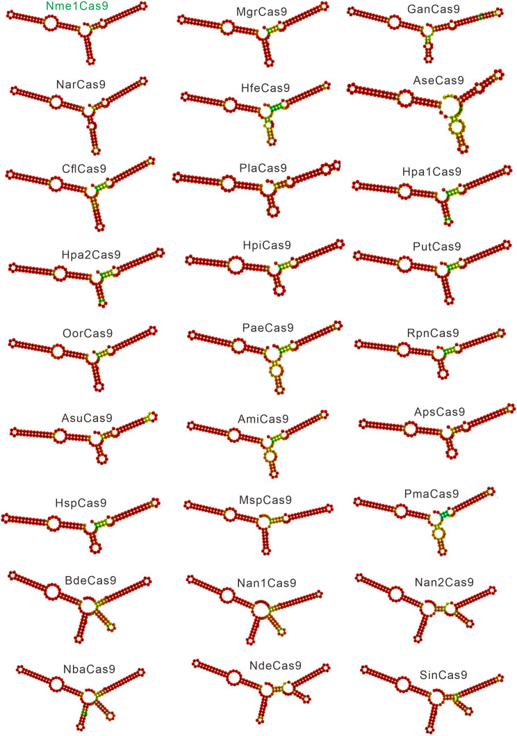
Figure 1—figure supplement 4. Single-guide RNA (sgRNA) scaffolds of Nme1Cas9 orthologs.

Figure 1—figure supplement 4.

In silico co-folding of the crRNA direct repeat and putative tracrRNA shows stable secondary structure and complementarity between the two RNAs.
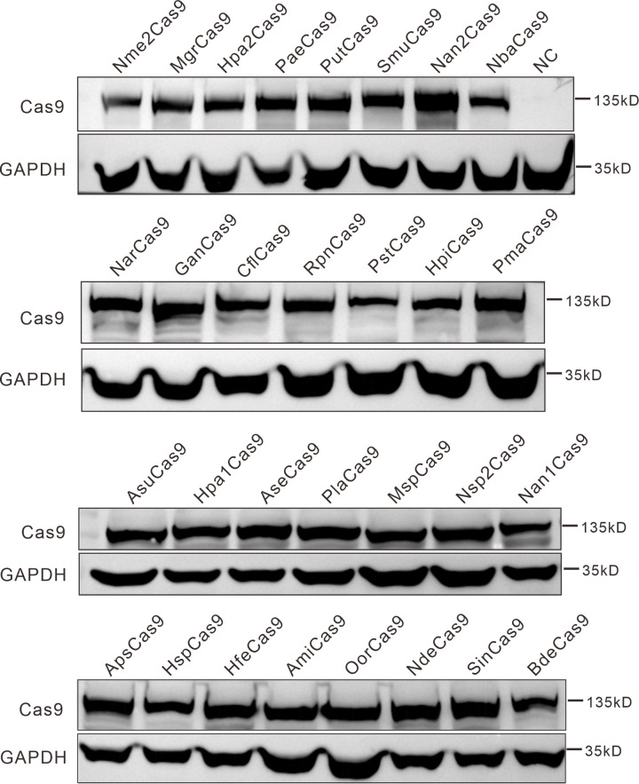
Figure 1—figure supplement 5. Protein expression levels of Nme2Cas9 orthologs were analyzed by western blot.

Figure 1—figure supplement 5.

HEK293T cells without Cas9 transfection were used as a negative control (NC).
Figure 1—figure supplement 5—source data 1. Source data for Figure 1—figure supplement 5.