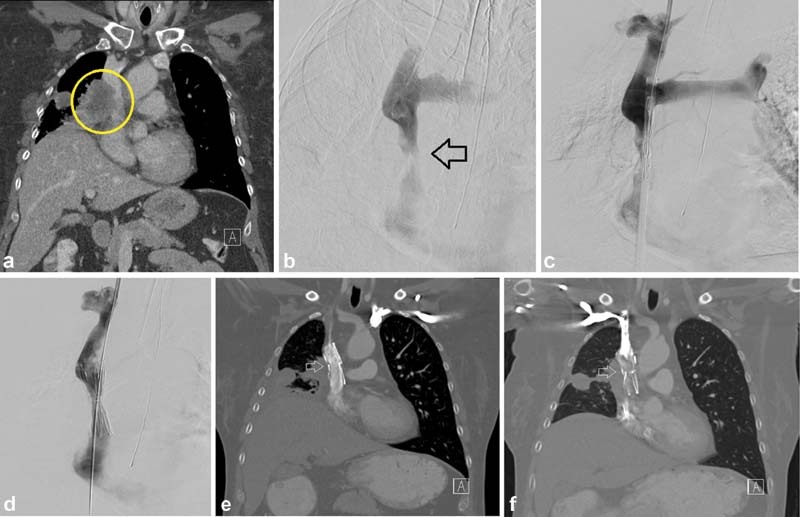
Fig. 7.

A 54-year-old woman presented with face, arm, and neck swelling as well as headaches. A CT showed large right perihilar tumor invading into the mediastinum and tumor invasion into the SVC (circle, a ), with a subsequent pathologic diagnosis of small cell lung cancer. Initial venogram and pre–stent deployment venogram ( b and c ) confirmed the SVC stenosis (arrow, b ). A 20-mm-diameter Cook-Z (Cook Medical; Bloomington, IN) stent was placed and post dilated to 10 mm ( d ). After tissue diagnosis was made, she was started on chemotherapy and radiation. CT 6 months post stenting showed a widely patent stent (arrow, e ). At 18 months, CT showed tumor invasion into the stent and SVC (arrow, f ).
