
Fig. 3.
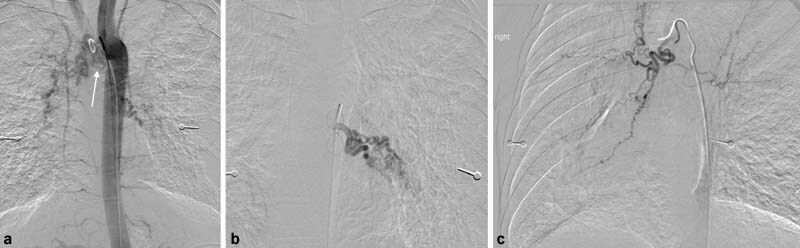
( a ) Flush thoracic aortogram demonstrates bilateral hypertrophied bronchial arteries in a 25-year-old man with cystic fibrosis and massive hemoptysis. Note the right bronchial artery has an ectopic origin from the underside of the aortic arch (arrow). ( b ) Selective angiogram of the abnormal, hypertrophied left bronchial artery. ( c ) Selective angiogram of the ectopic right bronchial artery shows a dilated, ectatic vessel with hyperemia.