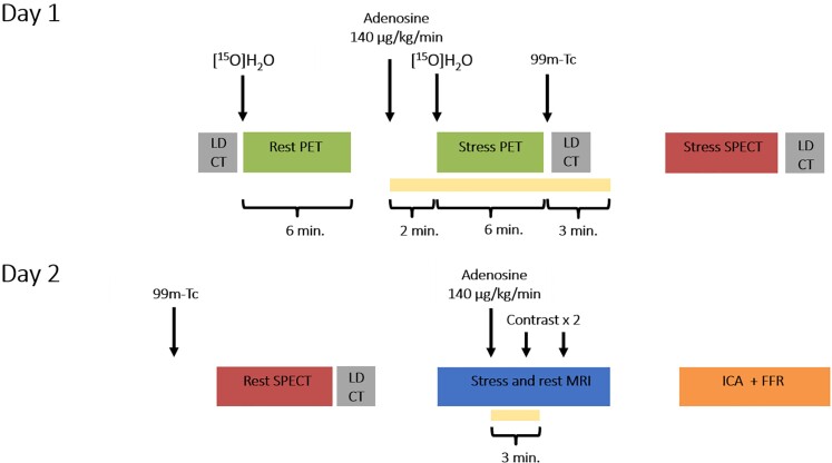
Figure 1.
Schematic illustration of the study protocol. All patients underwent the same 2-day protocol with positron emission tomography, single-photon emission computed tomography, and magnetic resonance imaging, followed by invasive coronary angiography with routine fractional flow reserve measurements. FFR, fractional flow reserve; ICA, invasive coronary angiography; LD-CT, low-dose computed tomography; MRI, magnetic resonance imaging; PET, positron emission tomography; and SPECT, single-photon emission computed tomography.

