
Fig. 7. Schematic of BAK activation by the 7D10 antibody.
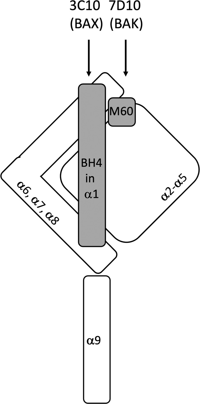
Non-activated BAK is shown with α9 inserted in the mitochondrial outer membrane. The α2-α5 core and α6-α8 latch are stabilised by contacts with both the BH4 domain (34VFRSYV39 in α1) and the M60 residue (in α1-α2 loop). The 7D10 antibody binds to 51GVAAPAD57 residues in the α1-α2 loop to trigger activation by disrupting M60’s linchpin role. In mitochondrial BAX the 3C10 antibody binds to a similar region, but triggers activation by disrupting contact with the BH4 domain [13].