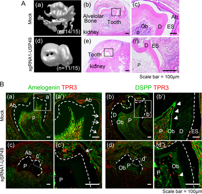
Fig. 8. Depletion of USP49 impairs the tooth development.
A Calcified teeth obtained after kidney capsule transplantation for 4 weeks. Three-dimensional (3D) reconstruction images from micro-computed tomography of calcified teeth in the (a) mock-group and (d) USP49-depletion group. H&E staining images from the (b, c) mock-group and (e, f) USP49-depletion group. Scale bar: 100 µm. (a (a, b)) In mock-group, the tooth is well calcified and surrounded by bone. (c) Pulp, odontoblasts, dentin, and ameloblasts were observed in the mock group. (a (d, e)) In the USP49-depletion group, the tooth is not fully calcified and not surrounded by bone. (f) Tooth showed defects in the pulp, dentin formation with few odontoblasts, and enamel formation with few ameloblasts. Fifteen biological replicates per group (n = 15) (H&E staining was performed from six biological replicates with randomly selected sections per group). B Amelogenin and DSPP expression levels in the (a and a’; b and b’) mock group and (c and c’, d and d’) USP49-depletion group. Scale bar = 100 µm. a’, b’, c’, and d’ are higher magnification images of a, b, c, and d, respectively. Ab, ameloblast; ES, enamel space; D, dentin; Ob, odontoblast; P, pulp; dotted line, dentinoenamel junction; arrows, positive cells in ameloblasts; arrow heads, positive cells in odontoblasts. The microscopic images from six biological replicates with randomly selected sections per group.

