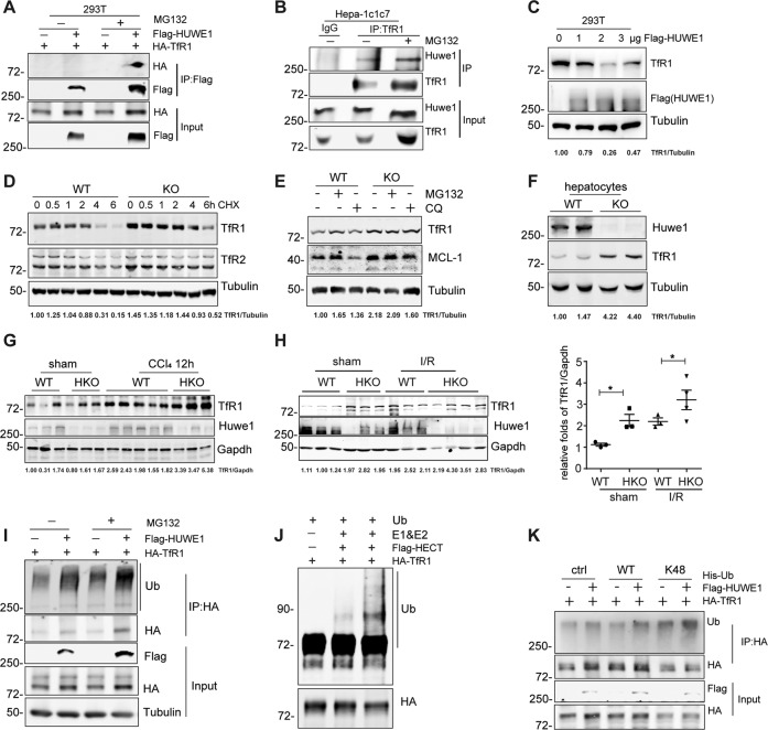
Fig. 5. HUWE1 ubiquitinates and degrades TfR1.
A Co-immunoprecipitation of HA-TfR1 with Flag-HUWE1 in HEK293T cells. Exogenous HUWE1 was immunoprecipitated using Flag-M2 beads, and the immunoprecipitants were immunoblotted with HA antibody. B Co-immunoprecipitation of Huwe1 with TfR1 in mouse liver hepatoma Hepa-1c1c7 cells. Endogenous TfR1 was immunoprecipitated using anti-TfR1, and the immunoprecipitants were immunoblotted with Huwe1 antibody. IgG, immunoglobulin G. C 293T cells were transfected with increasing levels of Flag-HUWE1, endogenous TfR1 was detected by western blot. Huwe1 WT and KO MEFs were treated with 15 μg/ml cycloheximide (CHX) (D), 10 µM MG132 or 10 µM Chloroquine (CQ) (E), endogenous TfR1 was immunoblotted. Mcl-1 was used as a positive control. F Immunoblotting of Huwe1 and TfR1 in hepatocytes isolated from Huwe1 WT and KO mice. G Hepatic Huwe1 and TfR1 were immunoblotted in Huwe1 WT and HKO mice challenged by CCl4 for 12 h. (H) Hepatic TfR1 were detected by immunoblotting in Huwe1 WT and HKO mice subjected to I/R for 6 h. TfR1 protein levels were normalized to those of Gapdh. *p < 0.05, compared in the indicated groups. I Ubiquitination of HA-TfR1 in HEK293T cells co-expressed with empty vector or Flag-HUWE1 treated by MG132 for 4 h before harvesting. J Recombinant TfR1 (100 nM) was incubated with ATP regenerating buffer, E1, E2, recombinant proteins of His tagged HECT domain of HUWE1 (10 nM, or 40 nM), and ubiquitin in a 30 μl reaction volume for 1 h at 37 °C. Three independent experiments were performed to confirm the ubiquitination of TfR1 by HUWE1 in vitro. K Immunoprecipitation analysis from HEK293T cells transiently co-transfected with HA-TfR1, Flag-HUWE1 and Wildtype or K48-Ubiquitin. The TfR1 bands in C–H were quantified with the Image J software, and the ratio of TfR1 to Tubulin or Gapdh in each lane was indicated on the bottom.

