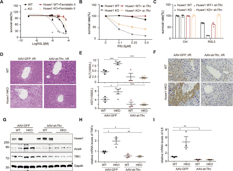
Fig. 7. Huwe1-mediated degradation of TfR1 mitigates ferroptosis and acute liver injury.
A, B TfR1 inhibition protected against Huwe1 KO sensitized ferroptotic cell death. Huwe1 WT and KO MEFs were pretreated with Ferristatin II for 4 h (A) or transfected with siRNA against Tfrc for 36 h (B), then subject to different concentrations of RSL3 treatment. Cell viability was evaluated with CellTiter Glo. C TfR1 was stably knocked down by shRNA in Huwe1 wildtype and knockdown Hepa-1c1c7 cells and then treated with RSL3. Cell viability was evaluated with CellTiter-Glo. All the data are representative of at least three independent experiments and presented as the means ± SD. D–I AAV9-shTfrc and AAV9-GFP were injected in the lateral tail vein of Huwe1 HKO and control mice to knock down Tfrc for 4 weeks, then mice were subjected to I/R surgery. Representative H&E staining images (D), serum ALT and AST levels (E), 4-HNE staining in the liver sections (F), and Acsl4 expression (G) were evaluated 6 h after I/R injury in the indicated groups. n = 3–5. Scale bars, 50 μm. Levels of cytokines (TNFα, IL6) in liver lysates were analyzed by real-time quantitative PCR (H–I, n = 3–4). Data and error bars are mean ± s.d., n = 3 independent repeats in A–C. *p < 0.05, **p < 0.01, compared in the indicated groups using unpaired two-tailed Student’s t test.

