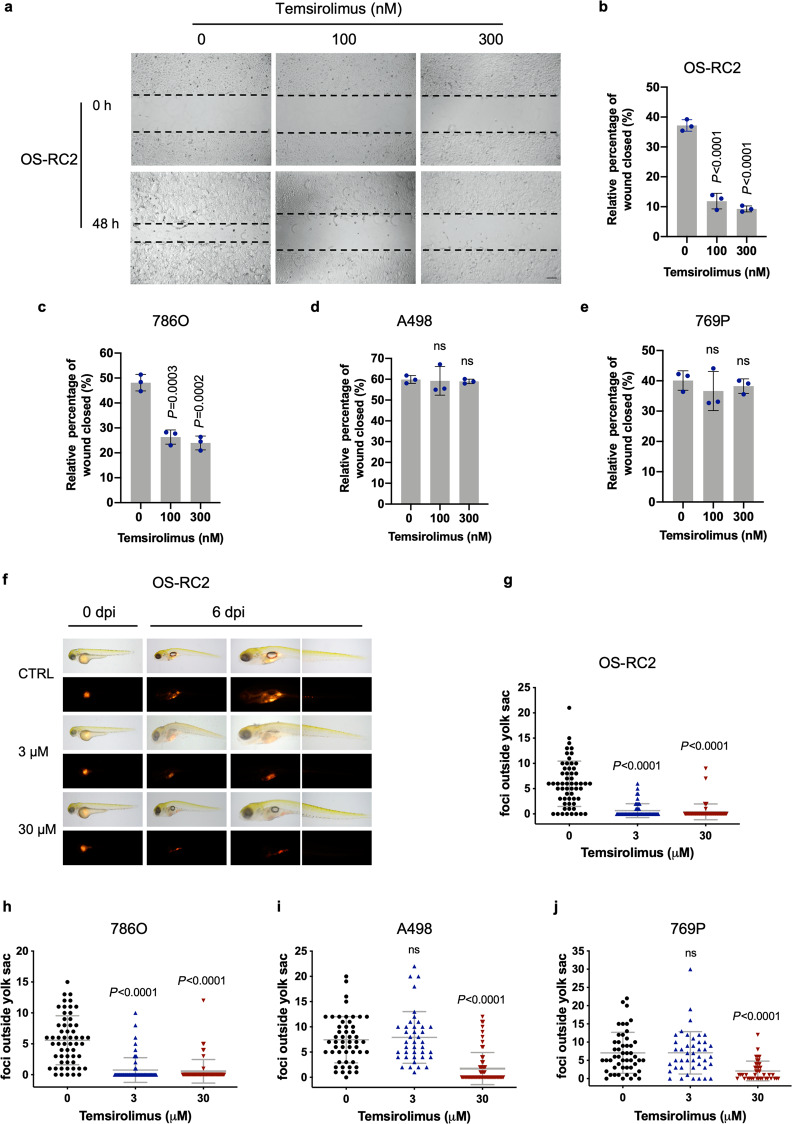
Fig. 5. Temsirolimus exhibits a more potent inhibitory effect on migration in PTEN-deficient ccRCCs than in PTEN-proficient ccRCC cells.
a Wound healing assays were performed to examine the effects of temsirolimus on the migration of OS-RC2 cells. Representative pictures of OS-RC2 cells at 0 h (time of wounding) and after 48 h of exposure to the indicated concentrations of temsirolimus. Scale bar, 200 μm. Quantification of wound healing, shown as the wound closure percentage in OS-RC2 (b), 786O (c), A498 (d), and 769P (e) cells. The data are presented as the mean ± SD (n = 3, five independent fields for each sample) of one representative experiment out of three. Significance was assessed using ANOVA followed by Tukey’s test. f Representative images of zebrafish embryos engrafted with OS-RC2 cells at 0 and 6 days post injection (dpi). Zebrafish embryos were treated with temsirolimus at the indicated concentrations. Quantification of the dissemination of OS-RC2 (g), 786O (h), A498 (i), and 769P (j) cells. Each dot represents the number of foci outside the yolk sac in a single embryo. The mean ± SD of three independent experiments is shown. Significance was assessed using ANOVA followed by Tukey’s test.

