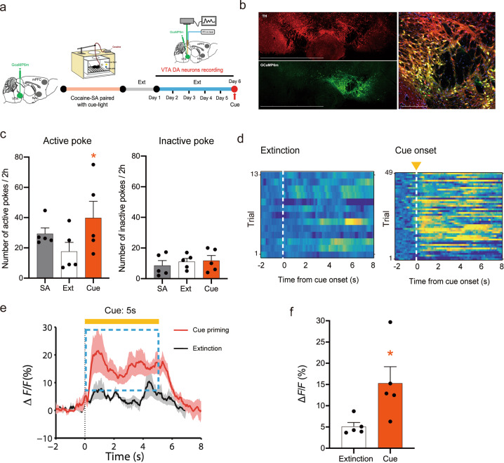
Fig. 4. Re-exposure to cocaine-associated cues activates VTA DA neurons.
a Schedule of the fiber photometry of VTA DA neurons in cue-induced cocaine seeking. b Representative images illustrating GCaMP6m expression in the VTA in DAT-Cre mice after intra-VTA AAV-GCaMP6m microinjection. 10×, scale bar = 1500 μm; 20×, scale bar = 200 μm. c Mean numbers of nose pokes in active and inactive holes during the last 3 sessions of cocaine self-administration (SA), the last 3 sessions of extinction, and the cue reinstatement test. SA-associated cue lights reinstated active-poke responses in mice after the extinction of SA. One-way RM ANOVA revealed a cue priming main effect: F(1,4) = 18.878, P = 0.012. Post hoc Bonferroni tests revealed that the differences in cue-induced active-poke responses were statistically significant (*P < 0.05, compared to the last session of extinction, n = 5). Inactive pokes were not affected by cue light re-exposure after the extinction of SA. One-way RM ANOVA revealed a cue priming main effect: F(1,4) = 0.0437, P = 0.845, n = 5. d The ΔF/F in cue priming and extinction are presented as heatmaps. Activation is presented in yellow, and depression is presented in blue. e The mean ΔF/F during the presence and absence of the cue (5 s) in extinction. The mean ΔF/F significantly increased during cue priming compared to the control condition in extinction, assessed by a paired t test, t = −2.552, *P < 0.05. f Quantification of the activity of VTA DA neurons (ΔF/F) in extinction and cue priming.

