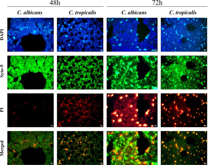
Figure 2.
Illustration of the biofilms of C. albicans and C. tropicalis at 48 and 72 h of growth by epifluorescence microscopy using 4′,6-diamidino-2-phenylindole fluorescent stain and LIVE/DEAD Biofilm Viability Kit. Time samples of 48 and 72 h were used to compare the total cell and live/dead cells in the biofilms using an Olympus BX50 microscope, and pictures were obtained by AmScope software at ×100 magnification. Then, the pictures from each filter were merged in Fiji-ImageJ version 1.57 (Schindelin et al., 2012).

