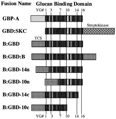
FIG. 1.
Schematic representation of fusion proteins containing a GBD. The positions of individual YG repeats (small hatched boxes) were kept constant in the figure. Numbers indicate how many repeats were still present in the truncated protein, and “n” or “c” indicates the terminus where the deletion occurred. GbpA carries its native N-terminal domain, while GBD-SKC contains the enzyme SKC at the C terminus of the GBD. All other fusion proteins were made with TCS and GBD. B-GB and B-GD (not shown) were similar to B-GBD, except that the GBD was derived from GTF-I and GTF-S, respectively.

