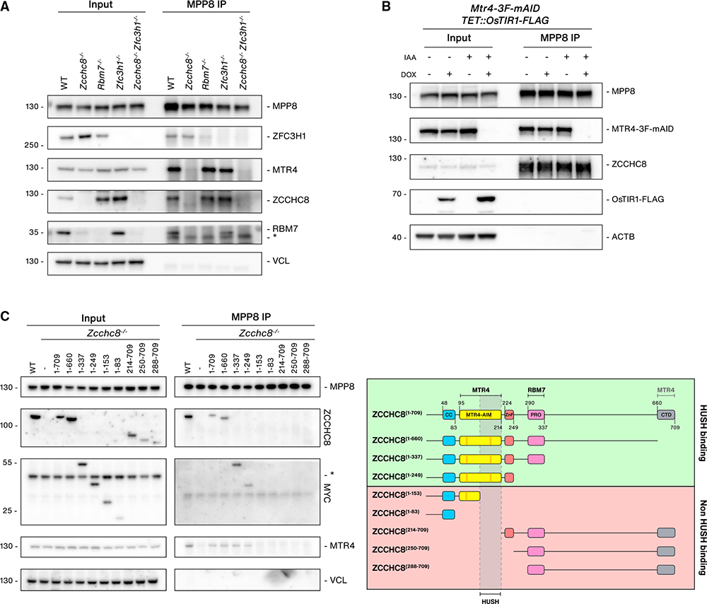
Figure 3. ZCCHC8 bridges the interaction between NEXT and HUSH.
(A) WB analysis of MPP8 IPs from lysates of WT, Zcchc8−/−, Rbm7−/−, Zfc3h1−/−, and Zcchc8−/−Zfc3h1−/− cells. Input and IP samples were probed with antibodies against HUSH-, NEXT-, and PAXT-related proteins as indicated and Vinculin (VCL, input loading control). Non-specific bands are indicated with an asterisk (*).
(B) WB analysis of MPP8 IPs from TET::OsTIR1-FLAG, MTR4-3F-mAID cells following doxycycline (DOX) and/or IAA treatment (4 h) as indicated. Input and IP samples were probed with antibodies against MPP8, MTR4, ZCCHC8 FLAG, and Actin (ACTB, input loading control).
(C) Left: WB analysis of MPP8 IPs from WT or Zcchc8−/− cells stably expressing MYC-tagged ZCCHC8 fragments labeled with amino acid numbers as in the right panel. Input and IP samples were probed with antibodies against MPP8, ZCCHC8, MYC, MTR4, and Vinculin (VCL, input loading control). Right: schematic representation of ZCCHC8 domains, generated fragments, and MPP8 IP data summary. Known protein binding regions are indicated on the top. Fragments shown to be HUSH binding (green) or not (red) are indicated, and a putative binding region is shown.

