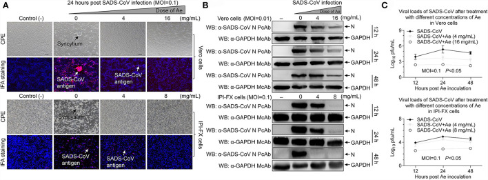
Figure 1.
The effect of Ae on SADS-CoV infectivity. (A) Vero and IPI-FX cells were pre-incubated with different concentrations of Ae (2–16 mg/ml) for 1 h, followed by infection with SADS-CoV at an MOI of 0.1. After 1.5 h, the cells were re-treated with Ae or the normal medium. The positive red IFA signals for SADS-CoV N protein were monitored by fluorescence microscopy at 24 hpi. CPE and SADS-CoV antigens were indicated by arrows. Vero and IPI-FX cells were treated as above described, cell lysates were prepared at indicated time points (12 h, 24 h, and 48 h), and the expression level of N and GAPDH proteins was examined by Western blot using anti-SADS-CoV N polyclonal antibody and anti-GAPDH monoclonal antibody (B), or viral titers were determined by the TCID50 assay (C). Results are representative of three independent experiments (mean ± SD). n = 3.

