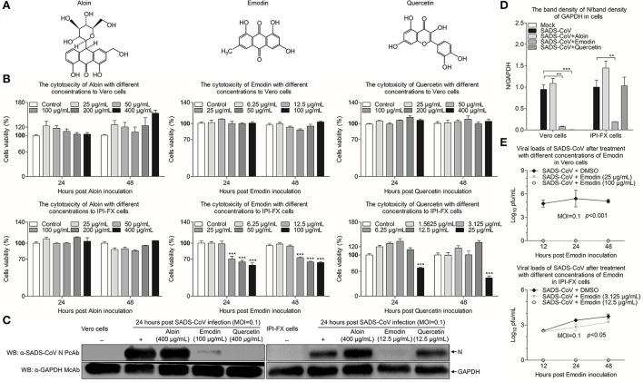
Figure 2.
Emodin from Ae inhibits SADS-CoV replication in vitro. (A) Chemical structures of Aloin, Emodin, and Quercetin. (B) The cytotoxicity of Aloin, Emodin, and Quercetin in Vero and IPI-FX cells. Vero and IPI-FX cells were co-incubated with various concentrations of Aloin (25–400 μg/ml), Emodin (6.25–100 μg/ml), and Quercetin (1.5625–400 μg/ml) or 0.1% DMSO for 24 h and 48 h prior to the CCK-8 assay. (C) Vero and IPI-FX cells were pre-incubated with Aloin, Emodin, and Quercetin, or 0.1% DMSO at a safe concentration for 1 h, followed by infection with SADS-CoV at an MOI of 0.1. After 1.5 h, the cells were re-treated with Aloin, Emodin, and Quercetin or the normal medium. At 24 h post-inoculation, the expression levels of N and GAPDH proteins in the cell lysates were detected by Western blot using anti-SADS-CoV N polyclonal antibody and anti-GAPDH monoclonal antibody. (D) The relative quantity of SADS-CoV N protein described in (C). (E) At indicated time points (12 h, 24 h, and 48 h), the viral titers in the cell lysates were determined by the TCID50 assay. Results are representative of three independent experiments (mean ± SD). n = 8 or 3. **p < 0.01, ***p < 0.001.

